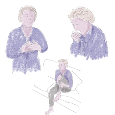
Patients with systemic mastocytosis (SM) report a substantial delay in diagnosis1
7YEARS
Median time from symptom onset to diagnosis1*
In that time, patients may present to a diverse group of specialists, including2:
- Allergists/immunologists
- Gastroenterologists
- Endocrinologists
- Primary care practitioners
- Dermatologists
- Haematologists/oncologists
Recognising common symptoms may help with earlier identification of SM
Skin lesions, anaphylaxis, diarrhoea?2
Consider SM
Identifying SM shouldn’t be a guessing game
Symptoms of SM can mimic other disorders
1. Other MCAS
MCAS3,4
Bone marrow biopsies for patients with MCAS may show 1 or 2 minor criteria for SM but do not fulfill the complete WHO criteria
2. Conditions in SM
Urticaria3
Urticaria pigmentosa is thought to be a benign, self-resolving condition. Unlike forms of mastocytosis, there is rarely any internal organ involvement, as would be found in patients with SM
Myeloproliferation3,5
At least 60–80% of advanced SM patients present alongside a neoplasm (CMML, MPN, MDS)
Malabsorption6
GI symptoms are present in 60–80% of SM patients. SM-related GI symptoms are largely caused by release of mediators and in rare, advanced forms by mast cell infiltration of the gut causing malabsorption
3. Other disorders that mimic SM -> differential diagnosis
CM3,4,7,8
The skin is the only site of involvement in cutaneous mastocytosis, whereas SM affects other organs besides skin
97% of patients presenting with adult-onset CM actually have SM†
Diagnosis of CM requires a bone marrow assessment, which may aid in confirming or excluding SM in the next step
IBD/IBS3,6
SM can cause GI symptoms similar to IBD
Endocrine disorders3,9
Although endocrine disorders can cause flushing, recurrent, unexplained flushing should raise a high index of suspicion for SM
*Based on data from 149 patients with self-reported mastocytosis in Mast Cell Connect registry in Jennings 2018 study.1
†Based on Berezowska 2014 study with 59 patients with the clinical diagnosis of adult-onset mastocytosis in the skin established between 2004 and 2008.7
CM=cutaneous mastocytosis; CMML=chronic myelomonocytic leukaemia; GI=gastrointestinal; IBD=inflammatory bowel disease; IBS=irritable bowel syndrome;
MCAS=mast cell activation syndrome; MDS=myelodysplastic syndrome; MPN=myeloproliferative neoplasm.
Recognising severe and recurrent instances of the common symptoms can raise the suspicion of SM: mast cell examination, serum tryptase assessment or high-sensitivity KIT D816V mutation testing can help diagnose SM2,8
Explore case studies of the types of patients you may see in your practice
The profiles below are examples of different patient types, which may help you recognise patients in your practice who may be at risk of SM. Patient profiles are fictionalised through a review of published literature and are not actual patients
Select each of the cases below to learn more

Victor,
age 67
Presenting to his
haematologist/oncologist
Victor:
SM-AHN confirmed on second evaluation
Patient history and presentation:
- Initial signs and symptoms and pathology work-up confirmed Victor’s diagnosis of chronic myelomonocytic leukaemia (CMML). At the time of CMML diagnosis, next-generation sequencing (NGS) on bone marrow detected TET2, SRSF2, RUNX1 and KIT D816V mutations
- After initiating first-line therapy with a hypomethylating agent, the patient returned for follow-up due to:
- unresolved splenomegaly
- recent onset of bone pain
- worsening fatigue
- unexplained weight loss
- increased episodes of diarrhoea
Full lab results:
- Complete blood count and chemistry profile normal
- Serum tryptase: persistently elevated, 110 ng/mL
- KIT D816V in bone marrow with high-sensitivity assay: reconfirmed positive mutation status
- IHC staining for CD25 and CD117 on bone marrow biopsy revealed CD25 spindle-shaped mast cells of >15 cells per cluster, >35% infiltration of cellularity by mast cells
Combined with the bone marrow mast cell aggregates, a confirmed KIT D816V mutation status allowed Victor’s diagnosis to be revised to SM with an associated haematological neoplasm (SM-AHN), where the -AHN component was CMML

Katherine,
age 42
Presenting to her
dermatologist
Katherine:
Indolent systemic mastocytosis presenting with skin lesions
Patient history and presentation:
- After presenting to her local dermatologist with bothersome maculopapular lesions on her torso and upper thigh, Katherine was diagnosed with cutaneous mastocytosis (CM)
- She was prescribed topical steroids for a year with limited relief
- Referred to allergist for second opinion after reporting recent onset of headaches, increasing fatigue and diarrhoea
- Had constant trouble focusing; expressed inability to work due to her symptoms. Reported increased frequency of ‘flare-ups’ and inadequacy of over-the-counter medications, such as H1, H2 blockers, in symptom management
- Lab results came back showing elevated serum tryptase at >100 ng/mL
Full lab results:
- Complete blood count, chemistry profile and liver function tests normal: overall stable organ function
- Follow-up serum tryptase: persistently elevated, 130 ng/mL
- KIT D816V in peripheral blood with high-sensitivity assay: positive mutation status
- Referred to haematologist/oncologist to conduct bone marrow biopsy. Pathology report revealed ~35% spindle-shaped bone marrow mast cells
With persistently elevated serum tryptase, >30% mast cell infiltration of bone marrow and a positive KIT D816V mutation status, Katherine’s diagnosis was revised to indolent systemic mastocytosis (ISM)
Victor:
SM-AHN confirmed on second evaluation
Patient history and presentation:
- Initial signs and symptoms and pathology work-up confirmed Victor’s diagnosis of chronic myelomonocytic leukaemia (CMML). At the time of CMML diagnosis, next-generation sequencing (NGS) on bone marrow detected TET2, SRSF2, RUNX1 and KIT D816V mutations
- After initiating first-line therapy with a hypomethylating agent, the patient returned for follow-up due to:
- unresolved splenomegaly
- recent onset of bone pain
- worsening fatigue
- unexplained weight loss
- increased episodes of diarrhoea
Full lab results:
- Complete blood count and chemistry profile normal
- Serum tryptase: persistently elevated, 110 ng/mL
- KIT D816V in bone marrow with high-sensitivity assay: reconfirmed positive mutation status
- IHC staining for CD25 and CD117 on bone marrow biopsy revealed CD25 spindle-shaped mast cells of >15 cells per cluster, >35% infiltration of cellularity by mast cells
Combined with the bone marrow mast cell aggregates, a confirmed KIT D816V mutation status allowed Victor’s diagnosis to be revised to SM with an associated haematological neoplasm (SM-AHN), where the -AHN component was CMML
Katherine:
Indolent systemic mastocytosis presenting with skin lesions
Patient history and presentation:
- After presenting to her local dermatologist with bothersome maculopapular lesions on her torso and upper thigh, Katherine was diagnosed with cutaneous mastocytosis (CM)
- She was prescribed topical steroids for a year with limited relief
- Referred to allergist for second opinion after reporting recent onset of headaches, increasing fatigue and diarrhoea
- Had constant trouble focusing; expressed inability to work due to her symptoms. Reported increased frequency of ‘flare-ups’ and inadequacy of over-the-counter medications, such as H1, H2 blockers, in symptom management
- Lab results came back showing elevated serum tryptase at >100 ng/mL
Full lab results:
- Complete blood count, chemistry profile and liver function tests normal: overall stable organ function
- Follow-up serum tryptase: persistently elevated, 130 ng/mL
- KIT D816V in peripheral blood with high-sensitivity assay: positive mutation status
- Referred to haematologist/oncologist to conduct bone marrow biopsy. Pathology report revealed ~35% spindle-shaped bone marrow mast cells
With persistently elevated serum tryptase, >30% mast cell infiltration of bone marrow and a positive KIT D816V mutation status, Katherine’s diagnosis was revised to indolent systemic mastocytosis (ISM)

Maria,
age 53
Presenting to ER
Maria:
ISM presenting with recurrent anaphylactic episodes
Patient history and presentation:
- Maria presented to ER after going into anaphylactic shock with hypotension from
a bee sting - Past medical history of chronic urticaria, managed with high-dose antihistamines
- Reported increased frequency of unexplained anaphylaxis (3 episodes in
previous month) - Recent onset of diarrhoea of daily occurrence
- Blood work revealed heightened risk for future anaphylactic episodes with elevated IgE and baseline serum tryptase (106 ng/mL)
- Referred to allergist on suspicion of underlying mast cell involvement
Full lab results:
- From allergist:
- Complete blood count and chemistry profile normal
- Serum tryptase: persistently elevated, 110 ng/mL
- Received MCAS diagnosis and referred to haematologist
- From haematologist: IHC staining for CD25 and CD117 on bone marrow biopsy revealed CD25 spindle-shaped mast cells of >15 cells per cluster, >35% infiltration of cellularity by mast cells; KIT D816V in peripheral blood with high-sensitivity assay: positive mutation status
With a positive KIT D816V mutation status, >35% infiltration of cellularity by mast cells, aberrant expression of CD25 and irregular, spindle-shaped mast cells, the haematologist was able to diagnose Maria with ISM

Peter,
age 55
Presenting to his
gastroenterologist
Peter:
ISM presenting with frequent diarrhoea and nausea
Patient history and presentation:
- Peter first presented to his gastroenterologist with diarrhoea, abdominal pain, nausea and weight loss
- Endoscopy showed inflammatory bowel disease managed with corticosteroids
- Peter returned to the gastroenterologist reporting increased frequency of diarrhoea and nausea that was preventing him from leaving the house
- Colonoscopy performed for further examination, revealing mucosal nodularity, prompting his gastroenterologist to take a sample for deeper analysis
- Histological sample revealed dense spindle-shaped mast cell infiltration of the lamina propria, showing involvement by mastocytosis
- Blood work that was taken aside colonoscopy showed elevated serum tryptase
(79.5 ng/mL) - Referred to haematologist on suspicion of underlying mast cell involvement
Full lab results:
- From gastroenterologist:
- Complete blood count and chemistry profile normal
- Serum tryptase: elevated, 79.5 ng/mL
- Referred to haematologist to conduct bone marrow biopsy
- From haematologist:
- IHC staining for CD25 and CD117 on bone marrow biopsy revealed CD25
spindle-shaped mast cells of >20 cells per cluster, >40% infiltration of cellularity by mast cells - KIT D816V in peripheral blood with high-sensitivity assay: positive mutation status
- IHC staining for CD25 and CD117 on bone marrow biopsy revealed CD25
With a positive KIT D816V mutation status, >40% infiltration of cellularity by mast cells, aberrant expression of CD25 and irregular, spindle-shaped mast cells, the haematologist was able to diagnose Peter with ISM
Maria:
ISM presenting with recurrent anaphylactic episodes
Patient history and presentation:
- Maria presented to ER after going into anaphylactic shock with hypotension from
a bee sting - Past medical history of chronic urticaria, managed with high-dose antihistamines
- Reported increased frequency of unexplained anaphylaxis (3 episodes in
previous month) - Recent onset of diarrhoea of daily occurrence
- Blood work revealed heightened risk for future anaphylactic episodes with elevated IgE and baseline serum tryptase (106 ng/mL)
- Referred to allergist on suspicion of underlying mast cell involvement
Full lab results:
- From allergist:
- Complete blood count and chemistry profile normal
- Serum tryptase: persistently elevated, 110 ng/mL
- Received MCAS diagnosis and referred to haematologist
- From haematologist: IHC staining for CD25 and CD117 on bone marrow biopsy revealed CD25 spindle-shaped mast cells of >15 cells per cluster, >35% infiltration of cellularity by mast cells; KIT D816V in peripheral blood with high-sensitivity assay: positive mutation status
With a positive KIT D816V mutation status, >35% infiltration of cellularity by mast cells, aberrant expression of CD25 and irregular, spindle-shaped mast cells, the haematologist was able to diagnose Maria with ISM
Peter:
ISM presenting with frequent diarrhoea and nausea
Patient history and presentation:
- Peter first presented to his gastroenterologist with diarrhoea, abdominal pain, nausea and weight loss
- Endoscopy showed inflammatory bowel disease managed with corticosteroids
- Peter returned to the gastroenterologist reporting increased frequency of diarrhoea and nausea that was preventing him from leaving the house
- Colonoscopy performed for further examination, revealing mucosal nodularity, prompting his gastroenterologist to take a sample for deeper analysis
- Histological sample revealed dense spindle-shaped mast cell infiltration of the lamina propria, showing involvement by mastocytosis
- Blood work that was taken aside colonoscopy showed elevated serum tryptase
(79.5 ng/mL) - Referred to haematologist on suspicion of underlying mast cell involvement
Full lab results:
- From gastroenterologist:
- Complete blood count and chemistry profile normal
- Serum tryptase: elevated, 79.5 ng/mL
- Referred to haematologist to conduct bone marrow biopsy
- From haematologist:
- IHC staining for CD25 and CD117 on bone marrow biopsy revealed CD25
spindle-shaped mast cells of >20 cells per cluster, >40% infiltration of cellularity by mast cells - KIT D816V in peripheral blood with high-sensitivity assay: positive mutation status
- IHC staining for CD25 and CD117 on bone marrow biopsy revealed CD25


